TAVI has become the treatment of choice for aortic stenosis in high-risk patients.
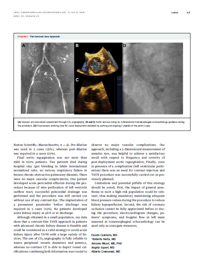
Among them, patients with pre-existing renal dysfunction are the most challenging ones to be treated: the use of even small amount of contrast dye could often deteriorate kidney function and badly impact on clinical outcome of these patients. proposed (2,3) but no one was able to completely abolish the use of contrast dye. We have recently introduced in our clinical practice a standardized protocol for performing transfemoral TAVI without contrast dye in patients with severe renal dysfunction (look at the img on the side). A no-contrast CT is usually performed in order to localize and quantify the degree of aortic calcification, the amount and the distribution of calcium in ilio-femoral vessels. Carbon dioxide angiography (angiography using CO2, which has basically no side effects for patients) is used in all patients with renal failure to measure vessel sizes and decide the suitability for TAVI the day before the procedure. In this setting, TAVI procedure is performed in general anaesthesi, with TEE guidance for annulus sizing. Our published data confirmed that this approach is feasible and effective, with no cases of acute kidney injury when using this novel and unique approach in very high-risk patients with GFR < 30 ml/min. In addition, the use of CO2 angiography is fully reliable to assess peripheral vessels diameters and patency, while no-contrast CT is able to depict vessel calcifications: combining both information was crucial to observe no major vascular complications.
We strongly believe that this strategy would become the default one in patients at high renal risk in selected and experienced centers.
Our approach

